
Poster abstracts
poster abstracts
1. Digitally encoded DNA nanostructures for multiplexed protein sensing with nanopores
Nicolas Bell, Ulrich Keyser
Cavendish Laboratory, University of Cambridge
2. Mechanosensitivity of the 2nd kind: TGFb mechanism of cell sensing the substrate stiffness
Max Cockerill, Matteo Escude, Michelle K. Rigozzi and Eugene M. Terentjev
Cavendish Laboratory, University of Cambridge
Cells can sense forces applied to them, but also the stiffness of their environment. These are two different phenomena, and here we investigate the mechanosensitivity of the 2nd kind: how the cell can measure an elastic modulus at a single point of adhesion, and how the cell can receive and interpret the chemical signal released from the sensor. Our model uses the example of large latent complex of TGFb as a sensor. Stochastic theory gives the rate of breaking of latent complex, which initiates the signalling feedback loop after the active TGFb release and leads to a change of cell phenotype driven by the a-smooth muscle actin. We analyse the timescale of approach to the steady state, the stability of the non-linear dynamical system, and how the steady-state concentrations of the key biomarkers vary depending on the elasticity of the substrate. We discover a crossover region for values of substrate elasticity closely corresponding to that of the fibroblast to myofibroblast transition. We suggest that the cell could actively vary the parameters of its dynamic feedback loop to `choose' the position of the transition region and the range of substrate elasticity that it can detect. The theory describes a variety of phenomena, such as the myofibroblast conversion in fibrosis of wounds and lungs and smooth muscle cell dysfunction in cardiac disease.
3. eScent® Context-Driven Wearable Scent Technology
Dr Jenny Tillotson
Institute of Biotechnology, Healthcare Biotechnology Group (Biosensors & Diagnostics), Department of Chemical Engineering & Biotechnology, University of Cambridge
eScent® introduces a new sensation and dynamic to the wearable technology sector. The sense of smell is our most primitive, and yet forgotten sense. eScent® offers an enabling platform technology and delivery device that emits fragrances at the right time, in the right place, depending on context. Embedded discreetly in "smart" jewellery and clothing, eScent® forms a localised 'scent bubble' around the face; an area of constant, detectable scent for the user based on biometric feedback, timer or pre-programmed from a smartphone. A spectrum of fragrances provides a new sensory awareness with wide‐ranging applications in fashion, fragrances for luxury brands, wellbeing, digital health, AR/VR, retail, sports, entertainment, learning and adult industry.
Fragrances are accurately targeted in short sharp bursts from a device based on biometric feedback, sound, or other analysis or sensor based trigger that is relevant to the situation, health condition or location, augmenting how we as humans interact with the physical world around us. This could be the release of counteractive wellbeing scents when stress levels are detected; the release of scents programmed to complement mood, music, gaming; or insect repellent released in response to the sound frequency of a mosquito.
With patents granted in UK, China and pending in the USA, eScent® has the potential to provide breakthrough progress in the fragrance industry; the advantage of sub-micron scent particles would be instant and sustained diffusion, depending on the user’s movements and surrounding conditions of air flow.
The application of miniaturization and novel biosensors to the latest fragrance development, linked with new insight into the power of fragrance ingredients to affect wellbeing, has the potential to change the whole way we appreciate, apply and use fragrances in the future.
4. Impedance-based biosensing on a disposable thin-film platform
Hanbin Ma, Yang Su, Chen Jiang, Nazrin Q. Azis and Arokia Nathan
Engineering Department, University of Cambridge
We present a disposable thin-film platform for impedance-based biosensing. Impedance-based measurements constitute a powerful tool for biosensing, since they provide the ability to identify both the physical and electrical properties of biomolecules. The measurement setup is less complicated compared to conventional biosensing techniques, based on optical- or mechanical-based sensors. The impedance sensor can be realised using the same material systems and processes used in large-area electronics, and the sensor can be disposable in view of its low fabrication costs. Large-area fabrication techniques also provide the flexibility to design and fabricate analyte-electronic interfaces (electrodes). With the help of finite element analysis, the electrodes geometry design can be optimised to achieve an enhanced sensitivity. Thin film fabrication techniques including both vacuum deposition and ink-jet printing also offer access to the application of a wide range of novel materials as the electrode interface. Some of the new low-dimensional materials show unique selectivity for biomolecules, which warrants further investigation. In addition, we have also integrated film-film electrodes into a conventional lab-on-a-chip system as a detector for micro-fluidic devices.
5. Design of an ATP sensor by using a fibre fabrication method
Jiyeong Chun, Jawad U. Rehman, and Elizabeth A. H. Hall
Department of Chemical Engineering and Biotechnology, University of Cambridge
To enhance the capacity of ATP detection, a new method of producing a platform embedding luciferase was developed. Luciferase, which has been fused to red fluorescent protein (Luc-RFP) retained its activity and affinity toward ATP in Luc-RFP solution. When the Luc-RFP fibre was extracted from air-water interface after 16 hours of film formation, the bioluminescence of luciferase was enhanced approximately 1.5 times compared to extracted fibre from Luc solution. Further scale-up of fibre fabrication was investigated through electrospinning, which can shorten the process of fibre fabrication to one hour. The mixture of Honeybee silk protein fused to RFP (RFP-HSP) and poly(vinyl alcohol) (PVA) was successfully electrospun in 10:1 weight ratio and produced a mat which consisted of uniform and smooth fibre in the diameter of 205 ± 23.3 um. The RFP-HSP mat was confirmed to retain its fluorescence even after the removal of PVA. It demonstrated that the functionality of protein is not affected by post-modification such as fibre extraction from air-water interface and electrospinning. This study suggests the possibility of fibre fabrication through electrospinning in the field of ATP sensing using Luc-RFP. Furthermore, it provides numerous biosensor applications using functional proteins fused to RFP.
6. Bio-inspired Immobilisation
Si Chen, Prof. Elizabeth A H Hall
Department of Chemical Engineering and Biotechnology, University of Cambridge
A challenge for amperometric biosensors is facilitation of immobilisation and directed electron transfer (DET). We explore the use of synthetic biology to engineer affinity polypeptide tags for redox enzymes, which can enable orientated enzyme immobilisation and thus, potentially shorten the electron transfer pathway for DET. mCherry and a flavin-containing redox enzyme, monomeric sarcosine oxidase (mSOx) were chosen as the model proteins to explore the concept. Polyhistidine (His) tag and silaffin (R5) tag were engineered to mSOx; both of the modified enzymes retained 100% of their enzymatic activity and were electrochemically active. His tag chelates to metal ions, therefore requires a metal binding ligand modification of the electrode for immobilisation. This only enables monolayer immobilisation of mSOx, so is expected to have limited success for enzymes with low catalytic activity. In contrast the R5 tag can precipitate silica in mild conditions, thus it can improve protein loading by entrapping the mCherry-R5 or mSOx-R5 within a silica matrix. It is shown that the enzyme's activity was retained within the silica matrix, with improved thermal. A suspension of carbon nanotubes (CNT) was incorporated into the enzyme entrapped silica matrix to enhance its conductivity, DET was observed when CNT incorporated silica matrix was adsorbed onto a suitable electrode.
7. Fast and Scalable Fabrication of ZnO Nanowires for Sensor Applications
Canlin Ou, Timothy Davies, Francesca Boughey, Pedro Sanchez-Jimenez, Sohini Kar-Narayan
Department of Materials Science and Metallurgy, University of Cambridge
The energy from ambient mechanical vibrations, like body movement, heartbeats, wind and fluid flow, may be directly transduced to electrical energy through the use of piezoelectric materials [1-2]. Piezoelectric sensors use the response of piezoelectric nanowires to vibrations for mechanical to electrical energy conversion. Using nanowires rather than bulk or thin films enhances electromechanical properties of the piezoelectric materials and they are more sensitive to low-amplitude vibrations [1], [3]. Most common piezoelectric materials are ceramic, but the brittle nature of these materials combined with poor reproducibility and little device optimisation make them not well suited to sensor applications. Here I report on a low temperature, cost effective and scalable novel fabrication techniques of ceramic-polymer nanocomposite into a small scale sensor. By using hydrothermal synthesis and electrodeposition techniques with nanoporous polymer membranes, an array of well-aligned ZnO nanowires was fabricated to form a flexible and robust sensor devices. Deformations of the device such as finger tapping, compression and bending could yield various electrical output signals depending on the applied forces and strains.
References
[1] Z. L. Wang et al., Piezoelectric nanogenerators based on zinc oxide nanowire arrays. Science. 312, 242-6 (2006).
[2] X. Wang, Piezoelectric nanogenerators – Harvesting ambient mechanical energy at the nanometer scale. Nano Energy. 1(1), 13-24, (2012).
[3] S. Crossley et al., Polymer-based nano-piezoelectric generators for energy harvesting applications. Mater. Sci. and Tech. (2014)
8. Flow imaging with absorption-based nonlinear tomography
Weiwei Cai and Clemens Kaminski
Department of Chemical Engineering, University of Cambridge
Optical imaging techniques are ubiquitous for the resolution of non-uniformities in gas flows. Planar imaging techniques such as laser-induced fluorescence have been well established and applied extensively in complicated turbulent reactive flows with both high temporal and spatial resolutions. However, the planar imaging suffers from one critical disadvantage, which is the requirement for continuous and abundant optical access that is sometimes not available for practical scenarios like engine tests. Tomography absorption spectroscopy (TAS) on the other hand, is highly complementary to planar imaging and can effectively reduce the demand for optical access using sparse sampling techniques. It has been proven so far the unique imaging possibility for in-cylinder/in-chamber engine measurements. TAS has been a mature technique for simultaneous imaging of temperature and species concentration, and become more versatile recently due to the progress in laser technology, spectroscopy, and theory of nonlinear tomography. The advancement in broadband frequency-agile laser sources massively enriches spectral information; and the nonlinear tomography enables the accommodation of multispectral information in a single tomographic inversion. The utilization of multispectral information improves the immunity of TAS to experimental noises and makes the simultaneous imaging of temperature, pressure, and multiple species possible. The nonlinear tomography also facilitates the imaging capacity of more sensitive and robust absorption techniques such as the calibration-free wavelength modulation spectroscopy even for optically dense measurements, extending the applicability of TAS to more general and harsh scenarios.
9. Hollow silica microspheres for combined sensing and delivery applications
Nadia Tsao, Dr. Jamie Walters, Professor Elizabeth Hall
Institute of Biotechnology, Department of Chemical Engineering and Biotechnology, University of Cambridge
Hollow silica microspheres have been demonstrated to be a good platform for sensing as well as a vehicle for drug delivery. Silica can be easily functionalised to contain reactive chemical groups for surface attachment of molecules while itself remaining chemically inert and biocompatible. The high surface area of mesoporous silica, coupled with the large inner volume, allows for the encapsulation of high concentrations of payload. In the case of an ion-sensing fluorescent payload, the thin nature of the silica shell significantly decreases the response time by moving from a spherical to a thin-layer geometry, while its micron size allows for easy detection by conventional microscopy. Hollow silica microspheres have also been shown to be easily internalized by cells through endocytosis, and are well adapted to the sustained delivery of cytotoxic drugs to cell culture. Here, a single hollow silica microsphere is proposed for combined sensing and delivery application to cell culture, where the effect of the drug delivery may be tracked directly in situ without further addition of reagent or processing.
10. Monitoring Tau Aggregation during Neuronal Transport Using a FRET sensor and Microfluidic-based culture chamber
Na Yu, Claire H. Michel, Clemens F. Kaminski, Gabriele S. Kaminski Schierle
Department of Chemical Engineering and Biotechnology, University of Cambridge
Alzheimer's disease (AD) is characterised by misfolding and aggregation of two amyloid proteins, amyloid β (Aβ) and Tau. Tau aggregates have recently been found to propagate from cell to cell in a prion-like manner. Hyperphosphorylation leads to its detachment from microtubules, which deconstructs the cell skeleton and disrupts axonal transport. To study these processes in detail we constructed a microfluidic device that permits us to investigate different neuronal compartments in isolation, and in particular to study trans-synaptic transfer of Tau aggregation. The device can be used with any optical microscopy technique, including optical nanoscopy methods. Here, we use a FRET sensor developed in our group to monitor aggregation reaction [1, 2, 3] and axonal transmission of tau in live differentiated SH-SY5Y cells via TCSPC-FLIM. In the device neurons were incubated with soluble K18-Tau labelled with Alexa 488 ('donor cells') and the subsequent uptake and aggregation was observed in wild type receiver cells ('acceptor cells'). Fluorescence lifetimes of K18-488 transmitted to the acceptor cells were found to be significantly lower than those in the donor cells, suggesting an acceleration of aggregation reactions on amyloid transmission, which may be significant in the context of disease. Furthermore, the transport takes only 20 min rather than 80 min upon neuronal stimulation with KCl.
References
[1] Kaminski-Schierle, G. S., et.al. 2011. Chemphyschem, 12, 673-80.
[2] Michel, C.H., et.al. 2014. J Biol Chem, vol. 289, pp. 956-67.
[3] Kaminski-Schierle, G. S., et.al. 2014. Bio-nanoimaging: Protein Misfolding & Aggregation, Academic Press, pp. 105-120
11. Characterization of mechanical properties of materials using Ultrasound Broadband Spectroscopy
Megha Agrawal, Abhinav Prasad, Prof J R Bellare, Dr A A Seshia
Nanoscience Center, Department of Engineering, University of Cambridge
This paper explores the characterization of homogenous materials (metals, alloys, glass and polymers) by a simple broadband ultrasonic interrogation method. The novelty lies in the use of ultrasound in a continuous way with very low input power (0 dBm or less) and analysis of the transmitted acoustic wave spectrum for material property characterization like speed of sound, density and dimensions of a material. Measurements were conducted on various thicknesses of samples immersed in liquid where continuous-wave, frequency swept ultrasonic energy was incident normal to the sample surface. The electro-acoustic transmission response is analyzed in the frequency domain with respect to a specifically constructed multi-layered analytical model. From the acoustic signature of the sample materials, material properties such as speed of sound and acoustic impedance can be calculated with experimentally derived values found to be in general agreement within 10% with the literature and with pulse-echo technique establishing the basis for a non-contact and non-destructive technique for material characterization. Further, by looking at the frequency spacing of the peaks of water when the sample is immersed, the thickness of the sample can be calculated independently from the acoustic response. This technique can prove to be an effective non-contact, non-destructive and fast material characterization technique for a wide variety of materials. A scaling analysis to motivate the basis for a MEMS platform is presented.
13. Red/Far-Red Optogenetics Tools for Systems Microscopy
Maximilian W. Fries, Kalina Haas, Emma K. Richardson, Alessandro Esposito and Ashok R. Venkitaraman
The Medical Research Council Cancer Unit, University of Cambridge
Over the last decade, traditional biochemical techniques have provided invaluable insights about the topology of biochemical networks, their dynamic behaviour and their correlation with cell fate [1]. However, state-of-the art biochemical tools – being applied to bulk measurements of proteins – cannot adequately quantify fast, asynchronous and heterogeneous biochemical signalling underlying cellular decisions [2]. Live cell fluorescence microscopy of individual cells expressing biosensors permits to overcome this limitation; however the number of sensors that can be read-out simultaneously is constrained by the broad emission spectra of fluorescent proteins. Recent developments in Optogenetics, now also permit us to manipulate biochemical activity with genetically encodable probes.
Here we describe the development of tools for a Systems Microscopy platform that allow multiplexing a number of FRET-based biosensors. We have developed novel FRET pairs, including the most red-shifted FRET pair that allows the efficient use of the full visible spectra. Moreover, the red/far-red spectral band can be used for the active control of biochemical network nodes with Optogenetics tools. Using a plant Phytochrome-based system, we demonstrate the control of oncogene signalling with light.
The integration of tools for quantitative parallel biochemical imaging and light-induced perturbation is likely to impact how biochemical (signalling or metabolic) networks are understood. Our developments represent a significant step forward in this direction.
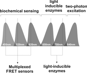
Illustration of the Systems Microscopy approach.
References
[1] S. D. M. Santos, P. J. Verveer, and P. I. H. Bastiaens, "Growth factor-induced MAPK network topology shapes Erk response determining PC-12 cell fate.," Nat. Cell Biol., 9, 324–330 (2007)
[2] A. Loewer and G. Lahav, "We are all individuals: causes and consequences of non-genetic heterogeneity in mammalian cells.," Curr. Opin. Genet. Dev., 21, 753–758 (2011)
14. Fabrication and Application of Porous Silicon for Optical Based Chemical and Biological Sensors
Paul Thomas Clarkson
Department of Chemistry and Department of Engineering, University of Cambridge
Porous silicon (PSi) represents a material that can be applied to the field of sensors. Its microporous structure, surface chemistry and vastly increased surface area-to-volume ratio make it an ideal surface to functionalise for sensing. The pre-established field of microelectronics and ready fabrication of silicon further supports the role that PSi can take in the future of sensors. This project combines PSi with optical waveguide sensors with the aim to functionalise the PSi waveguides for a new sensor platform.
15. Ellipsoid localisation microscopy infers the size and order of protein layers in Bacillus spore coats
James D. Manton, Julia Manetsberger, Miklós J. Erdélyi, Henry Lin, H. David Rees, Graham Christie, and Eric J. Rees
Department of Chemical Engineering and Biotechnology, University of Cambridge
Multi-layered protein coats are used for environmental protection, sensing, and interaction by many microorganisms, including spore-forming bacteria and viruses. In spores of Bacillus subtilis, over 70 distinct proteins make up a coat that is only about 100 nm thick, which helps keep the spore viable and able to germinate after a time as long as several decades in a harsh environment. Distinguishing the order of protein layers can indicate the function of different proteins – for example, which proteins form the outermost layers that protect the spore from lytic enzymes, and which hold the structure together? While fluorescent fusion proteins provide the only practical and non-invasive way to identify specific proteins in these multi-layered specimens, conventional optical microscopy lacks the resolution to resolve adjacent protein layers. Our original methods of fluorescent shell localisation make it possible to determine the order and geometry of concentric protein layers by fitting mathematical model structures to image data. Our method determines the radius of protein shells to a precision better than 10 nm, and produces layer orders for Bacillus subtilis and megaterium consistent with previous electron microscopy studies. In addition, the aspect ratio of elongated spores and the tendency of some proteins to localise near the poles can be quantified, enabling measurement of structural anisotropy. In future work, this technique will be used to optimise the structure of bacterial strains being developed for therapeutic drug delivery, in a joint project with MedImmune.
16. Film bulk acoustic resonators (FBAR) for sensing applications
M. DeMiguel-Ramos, G. Rughoobur, S. Esconjauregui, J. Robertson, E. Iborra and A. Flewitt
Electrical Engineering Department, University of Cambridge
Department of Electronic Engineering, Universidad Politecnica de Madrid
Gravimetric sensors can detect very small amounts of mass attached to their surface. Their operation principle is simple: when the targeted species binds to the surface of the sensor, its resonant frequency suffers a proportional shift to lower values. The higher the frequency, the higher the sensitivity. The most common example of this type of sensor is the Quartz Crystal Microbalance (QCM) widely used in vacuum technology and more recently in biosensing. However, quartz cannot be used in high frequency (above 300 MHz) due to its fragility and difficult handling. Film bulk acoustic wave resonator (FBAR) technology fills this gap and enables high frequency of operation and therefore higher sensitivity (up to the femtogram range). In this poster we present the advances in FBAR devices that the Electronic Devices and Materials group of the Engineering Department has achieved jointly with other groups around the world in the last years. Some examples include devices with two resonances for simultaneous measurement of mass and temperature, integration of carbon nanotubes in resonators to enhance sensitivity or in-liquid sensing using solidly mounted resonators.
17. Optical Sensors & Materials
Dominic J. Wales, Richard M. Parker, James C. Gates, Martin C. Grossel, Peter G.R. Smith, Andrew J.W. Physick, Julien Grand, Valeska P. Ting, Andrew D. Burrows, Svetlana Mintova, Chris R. Bowen
Department of Chemistry & Optoelectronics Research Centre, University of Southampton
Department of Chemical Engineering, University of Bath
Laboratoire Catalyse et Spectrochimie, ENSICAEN, Caen, France
The sensing of chemical species is required within a diverse set of fields including industry, environmental monitoring and homeland security. The sensing of gaseous chemicals has been traditionally achieved by use of electronic and electrochemical sensors. However, optical sensors demonstrate many benefits over these electronic systems, including remote interrogation of large sensor arrays via standard optical fibre and telecoms equipment and absence of spark risk in flammable environments.
I will present an overview of the recent work on optical sensing/detection of a range of chemical species. At the University of Southampton research has focused upon the use of supramolecular chemistry, surface chemistry and materials chemistry in combination with the integrated optical Bragg grating sensors. At the University of Bath and the Laboratoire Catalyse et Spectrochimie research has focused upon the development of porous materials (metal-organic frameworks and zeolites) as new sensing materials for exhaust gas sensing.
18. Photochemistry in optofluidic microreactors
Tijmen G. Euser
Cavendish Laboratory, Department of Physics, University of Cambridge
I am a new Lecturer in the NanoPhotonics Centre at the Cavendish Laboratory. Over the past years, our team at the Max Planck Institute for the Science of Light in Erlangen, Germany has demonstrated that hollow-core photonic crystal fibres act as excellent optofluidic waveguides that can be used to study highly-controlled light-matter interactions on sub-µL samples. The system enables highly-efficient photochemistry at optical powers a million times less than in conventional systems [1] and allows sensitive in-situ reaction monitoring, which is of great importance in catalytically active fibre [2].
Central to my research in Cambridge, I will establish an optofluidic microreactor laboratory in the Maxwell Centre, with the aim to develop novel methods for in-situ spectroscopy in these systems. These fundamental studies are expected to have a strong impact on the development of new catalytic and photochemical systems for advanced applications such as water-splitting, photo-chemical reduction of trace pollutants, and the development of novel light-activated anti-cancer drugs.
References
[1] A. M. Cubillas et al., "Photonic crystal fibres for chemical sensing and photochemistry", Chem. Soc. Rev. 42, 8629 (2013), front cover
[2] M. Schmidt et al. , "Chemical and (Photo)-Catalytical Transformations in Photonic Crystal Fibers", Chem. Cat. Chem. 5, 641 (2013), front cover
19. Investigation of dendritic spines by STED nanoscopy
Nathan Curry, Pierre Mahou, Janin Lautenschlaeger, Claire Michel, Laurie Young, Gabriele Kaminski-Schierle, Clemens Kaminski
Department of Chemical Engineering and Biotechnology, University of Cambridge
Parkinsons disease (PD) and dementia with Lewey bodies (DLB) are associated with the aggregation of proteins such as α-synuclein and amyloid-β. These proteins are thought to be linked to the loss of function of neuronal cells in these diseases, however it is not clear at what stage of aggregation these proteins interfere with cells.
One pathological hallmark of PD and DLB is the retraction of dendritic spines of neurons. To investigate which aggregation states have the greatest effect on cells we quantitatively measure the changes of length of dendritic spines in the presence of α-synuclein and amyloid-β at different stages of aggregation.
Dendritic spines range in length between 160-2000 nm so a quantitative investigation of spine length is challenging with conventional microscopes – whose resolution is limited to ~250 nm. Here we develop a STED super-resolution microscope to image dendritic spines beyond the diffraction limit.
We show the STED microscope which was developed for this application and demonstrate that it is capable of a resolution down to 40 nm on ideal samples and show that the SiR-actin dye can be used to stain dendritic spines and achieve super-resolved images.
Together this will allow a quantitative investigation of dendritic spine morphology and allow the investigation of the effects of aggregated proteins.
20. Dry mass sensing
T. Kartanas1, J. Charmet1, T. Muller1, R. Daly2 and T. P. J. Knowles1
1Department of Chemistry, University of Cambridge
2Department of Engineering, University of Cambridge
There is an urgent, yet currently unmet medical need for the development of accurate diagnostic tests for protein misfolding and aggregation diseases such as Alzheimer's disease and Parkinson's disease. Micro/nano-mechanical resonators have been identified as promising candidates for low cost, high resolution gravimetric biosensing. However, a common problem with current micro/nano-resonators based sensors is the device integration with microfluidics. Usually devices are functionalised with capture molecules and the devices are operated in liquid which leads to high dissipation. This results in low quality factors affecting the transducer frequency resolutions as well as other effects such as viscous drag that complicates the measurement interpretation. We are applying a completely orthogonal approach by detecting the dry mass of the molecules of interest in air. We have built a platform which allows uniform deposition of analytes on a microcantilever's surface using a microfluidic spray. The solvent evaporates quickly enabling measurement of the analytes dry mass. This configuration enables us to retain the high resolution associated with measurement in air as the damping losses are minimised.
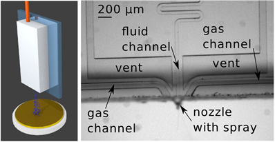
Dry mass sensing. The principle relies on spraying droplets onto a gravimetric sensor using microfluidics. As the solvent evaporates the dry solute mass can be measured thus minimizing damping losses. This configuration enables high resolution measurement.
21. Imaging Development on a Budget: A Novel Low-Cost Solution for Live Cell Imaging
Fergus Riche
Department of Engineering, University of Cambridge
The recent availability of ethically assured and biologically versatile sources of stem cells has made them an increasingly popular object of study in the fields of developmental biology and regenerative medicine alike. The development of the Yamanaka protocol for reversing the process of differentiation in adult cells has enabled not only the wider study of stem cell biology, but also the study of individual-focused medicine. In addition to this, the field of optical imaging has recently benefited from a number of advancements that have for the first time, made optical components and processing cheaper than mechanical solutions. In contrast to the ever increasing popularity of complex imaging modalities such as differential interference contrast, it has been found that the simplest of imaging modalities, brightfield illumination, could produce acceptable image quality for morphological analysis of cellular development when the images were processed sufficiently. A method for noise reduction of oversampled images is proposed, which was incorporated into the workflow of the imaging system, and validated by the production of good results from the analysis of images by an optical flow algorithm. Moreover, a quantitative test of resolving power is described, and used to compare the performance of the designed system to a commercial system used for cell observation, which shows similar results for the two systems despite their large differences in cost and size.
22. Multi-Channel FLIM-FRET: A Method for the Quantification of FRET Stoichiometry Using Phasor Plots and Acceptor Lifetime In-growth
WeiYue Chen, Edward Avezov, Simon C. Schlachter, Fabrice Gielen, Romain F. Laine, Heather P. Harding, Florian Hollfelder, David Ron, and Clemens F. Kaminski
Department of Chemical Engineering and Biotechnology, University of Cambridge
Förster resonance energy transfer (FRET) is a powerful tool for the study of protein-protein interactions in biological samples. The FRET efficiency, the stoichiometry of donors and acceptors, and the affinity between interacting molecules are all of interest for biochemical analyses using FRET. However, with conventional methods it is difficult to fully quantify these parameters due to donor fluorescence bleed-through and acceptor cross-excitation, which typically occur in FRET experiments. Here, we present a new method for the simultaneous quantification of the interaction parameters, including fractions of bound acceptors and donors, local protein concentrations and dissociation constants, in each image pixel, which we refer to as 'multi-channel FLIM-FRET (MC-FLIM-FRET)'. The method requires fluorescence lifetime imaging microscopy (FLIM) measurements in at most three different fluorescence channels, and makes use of phasor plot analysis. We demonstrate the capability of our method with microfluidic and cell experiments, and are able to quantify dissociation constants between tagged versions of glutathione (GSH) and glutathione S-transferase (GST), and the concentration of competitor reactants. The method is rigorously validated theoretically and with simulations.